Ectopic Molar in The Condylar Region: An Unusual Presentation

Impacted third molars are very common condition which we encounter in our daily practice. Ectopic teeth are those which are impacted in unusual positions or have been displaced from their normal anatomic locations. Ectopic mandibular third molars in the condylar region are rare and under reported. Most of the cases are seen in female patients with common signs and symptoms of pain, discomfort and swelling in the mandibular region or in the preauricular region with temperomandibular joint pain and discomfort. Here we report an asymptomatic case of ectopic third molar in the condylar region and follow up of the case with Cone Beam Computed Tomography (CBCT) imaging Keywords: Ectopic molar, Condylar region, impacted third molar, CBCT

By |2023-04-27T23:30:55+05:30April 27, 2023|Comments Off on Ectopic Molar in The Condylar Region: An Unusual Presentation

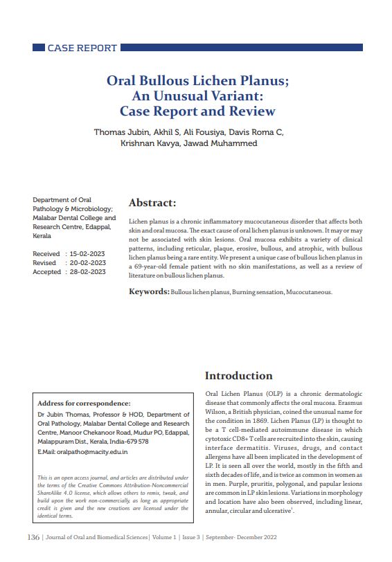
Oral Bullous Lichen Planus; An Unusual Variant: Case Report and Review

Lichen planus is a chronic inflammatory mucocutaneous disorder that affects both skin and oral mucosa. The exact cause of oral lichen planus is unknown. It may or may not be associated with skin lesions. Oral mucosa exhibits a variety of clinical patterns, including reticular, plaque, erosive, bullous, and atrophic, with bullous lichen planus being a rare entity. We present a unique case of bullous lichen planus in a 69-year-old female patient with no skin manifestations, as well as a review of literature on bullous lichen planus. Keywords: Bullous lichen planus, Burning sensation, Mucocutaneous

By |2023-05-12T11:45:56+05:30April 28, 2023|Comments Off on Oral Bullous Lichen Planus; An Unusual Variant: Case Report and Review

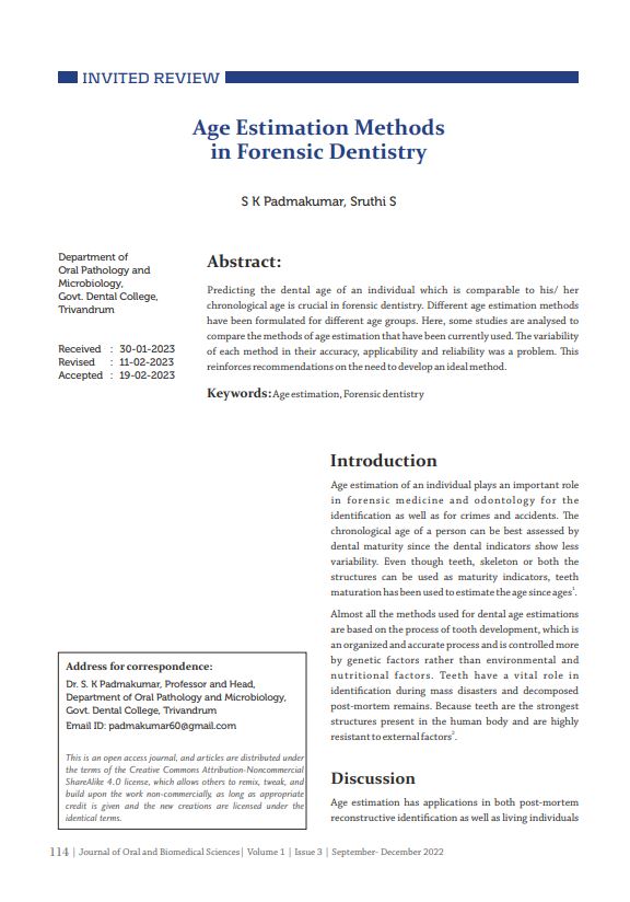
Age Estimation Methods in Forensic Dentistry

Predicting the dental age of an individual which is comparable to his/ her chronological age is crucial in forensic dentistry. Different age estimation methods have been formulated for different age groups. Here, some studies are analysed to compare the methods of age estimation that have been currently used.The variability of each method in their accuracy, applicability and reliability was a problem. This reinforces recommendations on the need to develop an ideal method. Keywords – Age estimation, Forensic dentistry

By |2023-05-12T11:48:58+05:30April 27, 2023|Comments Off on Age Estimation Methods in Forensic Dentistry

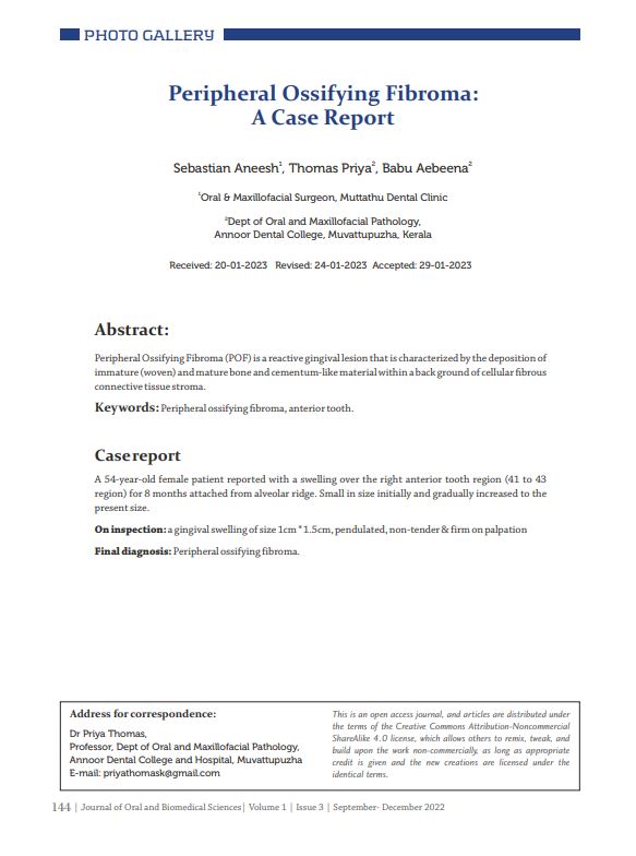
Peripheral Ossifying Fibroma: A Case Report

Peripheral Ossifying Fibroma (POF) is a reactive gingival lesion that is characterized by the deposition of immature (woven) and mature bone and cementum-like material within a back ground of cellular fibrous connective tissue stroma. Keywords: Peripheral ossifying fibroma, anterior tooth

By |2023-04-27T23:40:47+05:30April 27, 2023|Comments Off on Peripheral Ossifying Fibroma: A Case Report

A Rare Case of Osteolipoma Presenting as a Mobile Swelling on The Buccal Sulcus

Osteolipoma is an extremely rare histologic variant of lipoma that contains mature lamellar bone within the tumor and is independent of bone tissue. It is most commonly seen in buccal mucosa, floor of the mouth, tongue, palate, and parapharyngeal spaces of the oropharyngeal region. We present a case of 72 yr old female patient with a complaint of intraoral swelling in left lower buccal sulcus. The swelling was mobile and firm to hard in consistency. Excision specimen showed circumscribed mass of mature adipose tissue with metaplastic lamellated bone formation. The lesion was diagnosed as osteolipoma. The pathogenesis and the differential diagnosis are discussed with this case presentation. Keywords: - Lipoma, Osteolipoma, Buccal sulcus

By |2023-04-27T23:07:47+05:30April 27, 2023|Comments Off on A Rare Case of Osteolipoma Presenting as a Mobile Swelling on The Buccal Sulcus

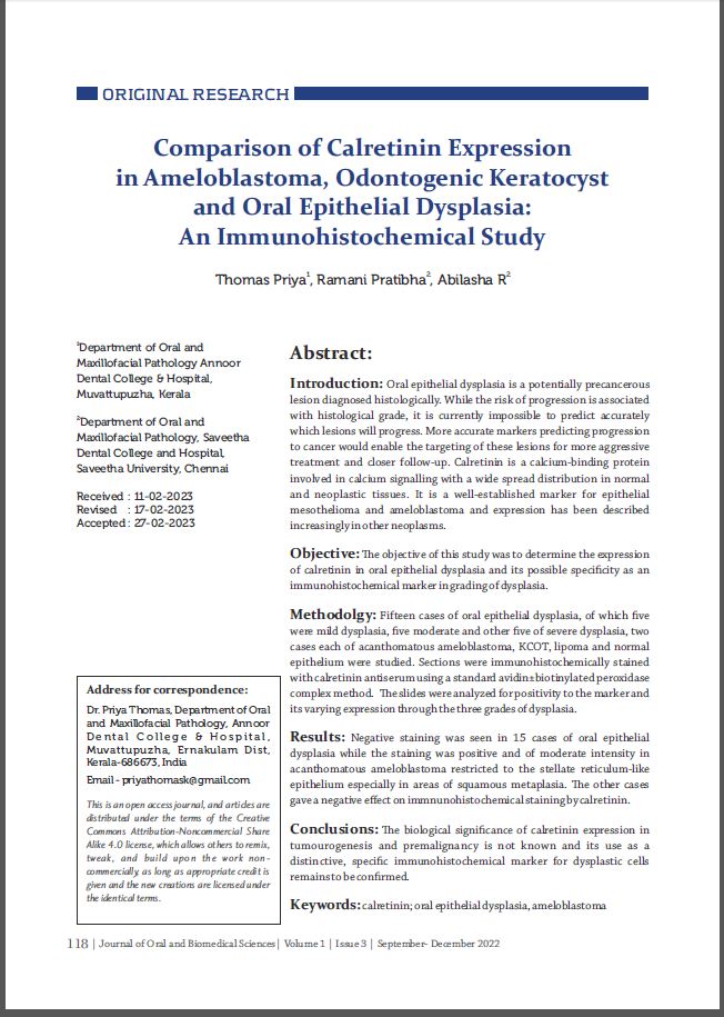
Comparison of Calretinin Expression in Ameloblastoma, Odontogenic Keratocyst and Oral Epithelial Dysplasia: An Immunohistochemical Study

Introduction: Oral epithelial dysplasia is a potentially precancerous lesion diagnosed histologically. While the risk of progression is associated with histological grade, it is currently impossible to predict accurately which lesions will progress. More accurate markers predicting progression to cancer would enable the targeting of these lesions for more aggressive treatment and closer follow-up. Calretinin is a calcium-binding protein involved in calcium signaling with a wide spread distribution in normal and neoplastic tissues. It is a well-established marker for epithelial mesothelioma and ameloblastoma and expression has been described increasingly in other neoplasms. Objective: The objective of this study was to determine the expression of calretinin in oral epithelial dysplasia and its possible specificity as an immunohistochemical marker in grading of dysplasia. Methodology: Fifteen cases of oral epithelial dysplasia, of which five were mild dysplasia, five moderate and other five of severe dysplasia, two cases each of acanthomatous ameloblastoma, KCOT, lipoma and normal epithelium were studied. Sections were immunohistochemically stained with calretinin antiserum using a standard avidin±biotinylated peroxidase complex method. The slides were analyzed for positivity to the marker and its varying expression through the three grades of dysplasia. Results: Negative staining was seen in 15 cases of oral epithelial dysplasia while the staining was positive and of moderate intensity in acanthomatous ameloblastoma restricted to the stellate reticulum-like epithelium especially in areas of squamous metaplasia. The other cases gave a negative effect on immnunohistochemical staining by calretinin. Conclusion: The biological significance of calretinin expression in tumourogenesis and premalignancy is not known and its use as a distinctive, specific immunohistochemical marker for dysplastic cells remains to be confirmed Keywords: calretinin; oral epithelial dysplasia, ameloblastoma

By |2023-05-12T11:44:03+05:30April 27, 2023|Comments Off on Comparison of Calretinin Expression in Ameloblastoma, Odontogenic Keratocyst and Oral Epithelial Dysplasia: An Immunohistochemical Study
Go to Top